Email Alert | RSS    帮助

中国防痨杂志 ›› 2020, Vol. 42 ›› Issue (3): 227-232.doi: 10.3969/j.issn.1000-6621.2020.03.009

• 论著 • 上一篇    下一篇

CT扫描对腹膜弥漫性病变的鉴别诊断价值

洪盟,过丽芳(),张建梅,王梦君,王仁贵   

  1. 100038 首都医科大学附属北京世纪坛医院放射中心
  • 收稿日期:2019-10-27 出版日期:2020-03-10 发布日期:2020-03-18
  • 通信作者: 过丽芳 E-mail:guolifang2008@sina.com

Value of CT scan for diagnosis and differential diagnosis of diffuse peritoneal lesions

HONG Meng,GUO Li-fang(),ZHANG Jian-mei,WANG Meng-jun,WANG Ren-gui   

  1. Department of Radiology,Beijing Shijitan Hospital,Capital Medical University,Beijing 100038,China
  • Received:2019-10-27 Online:2020-03-10 Published:2020-03-18
  • Contact: Li-fang GUO E-mail:guolifang2008@sina.com

摘要:

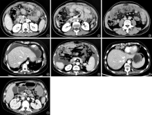
目的 分析3种比较常见腹膜弥漫性病变的CT扫描特征,探讨CT扫描对3种腹膜弥漫性病变的诊断与鉴别诊断价值。方法 回顾性分析首都医科大学附属北京世纪坛医院影像资料库中72例腹膜弥漫性病变患者资料,其中结核性腹膜炎16例(结核组),腹膜转移癌34例(转移癌组),腹膜间皮瘤22例(间皮瘤组)。比较3组患者的CT表现特征,以评价CT扫描对腹膜弥漫性病变的诊断与鉴别诊断价值。结果 (1)结核组、转移癌组与间皮瘤组壁腹膜均匀增厚的发生率分别为62.5%(10/16)、23.5%(8/34)和 27.3%(6/22),结核组与转移癌组、结核组与间皮瘤组比较,差异均有统计学意义(χ 2=5.221,P=0.022; χ 2=10.795,P=0.010);以上3组病变出现大网膜污垢样增厚的发生率分别为43.8%(7/16)、2.9%(1/34)和13.6%(3/22),结核组与转移癌组、结核组与间皮瘤组比较,差异均有统计学意义(χ 2=14.567,P=0.000; χ 2=4.332,P=0.037)。以上3组 “网膜饼征”的发生率分别为6.2%(1/16)、50.0%(17/34)和27.3%(6/22),结核组和转移癌组比较差异有统计学意义(χ 2=9.039,P=0.003);结核组与间皮瘤组比较差异无统计学意义(χ 2=1.216,P=0.270)。以上3组大网膜结节或肿块状增厚的发生率分别为12.5%(2/16)、14.7%(5/34)和50.0%(11/22),结核组与间皮瘤组、转移癌组与间皮瘤组比较,差异均有统计学意义(χ 2=5.788,P=0.016;χ 2=8.153,P=0.004)。(2)以上3组中少量腹腔积液的发生率分别为75.0%(12/16)、32.4%(11/34)和40.9%(9/22),出现大量腹腔积液的发生率分别为25.0%(4/16)、67.6%(23/34)和59.1%(13/22)。结核组与转移癌组、结核组与间皮瘤组出现中少量腹腔积液的发生率两组比较,差异均有统计学意义(χ 2=7.966,P=0.005;χ 2=4.354,P=0.037);结核组与转移癌组、结核组与间皮瘤组出现大量腹腔积液的发生率两组比较,差异均有统计学意义(χ 2=7.966,P=0.005;χ 2=4.354,P=0.037)。(3)以上3组出现心膈角淋巴结肿大的发生率分别为6.2%(1/16)、5.9%(2/34)和40.9%(9/22),间皮瘤组与结核组、间皮瘤组与转移癌组比较,差异均有统计学意义(χ 2=5.739,P=0.017;χ 2=10.382,P=0.001)。结论 3组患者在腹膜病变发生部位、形态、腹腔积液、淋巴结肿大等方面各体现了不同的CT特征, CT扫描对腹膜弥漫性病变进行鉴别诊断时具有重要价值。

关键词: 腹膜疾病, 腹膜炎, 结核性, 腹膜肿瘤, 体层摄影术, X线计算机, 诊断, 鉴别

Abstract:

Objective To analyze CT imaging characteristics of three common diffuse peritoneal lesions,and to explore the value of CT in the diagnosis and differential diagnosis of diffuse peritoneal lesions. Methods We retrospectively analyzed characteristics of CT imaging in 72 cases with diffuse peritoneal lesions, including 16 cases with tuberculous peritonitis (TBP), 34 cases with peritoneal metastasis (PM),22 cases with peritoneal mesothelioma (PMM). Results (1) The incidence rates of uniform peritoneal thickening were 62.5% (10/16) in TBP, 23.5% (8/34) in PM, 27.3% (6/22) in PMM, the differences between TBP and PM, TBP and PPM were statistically significant (χ 2=5.221,P=0.022; χ 2=10.795,P=0.010); the incidence rates of omentum majus dirt-like thickening were 43.8% (7/16) in TBP, 2.9% (1/34) in PM, 13.6% (3/22) in PMM, the differences between TBP and PM, TBP and PPM were statistically significant (χ 2=14.567,P=0.000; χ 2=4.332,P=0.037); the incidence rates of “omentum cake sign” were 6.2% (1/16) in TBP, 50.0% (17/34) in PM, 27.3% (6/22) in PMM, there was statistically significant difference between TBP and PM (χ 2=9.039,P=0.003), but difference was not statistically significant between TBP and PMM (χ 2=1.216,P=0.270); the incidence rates of nodules or mass thickening of greater omentum were 12.5% (2/16) in TBP, 14.7% (5/34) in PM, 50.0% (11/22) in PMM, the differences were statistically significant between TBP and PMM,PM and PMM (χ 2=5.788,P=0.016;χ 2=8.153,P=0.004). (2) The incidence rates of medium and small amount of ascitic fluid were 75.0% (12/16) in TBP, 32.4% (11/34) in PM, 40.9% (9/22) in PMM, there were statistically significant differences between TBP and PM,TBP and PMM (χ 2=7.966,P=0.005;χ 2=4.354,P=0.037); the incidence rates of large amount of ascitic fluid were 25.0% (4/16) in TBP, 67.6% (23/34) in PM, 59.1% (13/22) in PMM,there were statistically significant differences between TBP and PM,TBP and PMM (χ 2=7.966, P=0.005; χ 2=4.354, P=0.037). (3) The incidence rates of cardiac phrenic angle lymphadenopathy were 6.2% (1/16) in TBP, 5.9% (2/34) in PM, 40.9% (9/22) in PMM, the incidence differences between PMM and TBP, PMM and PM were statistically significant (χ 2=5.739,P=0.017;χ 2=10.382,P=0.001). Conclusion The specific visual characteristics on CT imaging, such as the location of peritoneal lesions, morphology, ascitic fluid and lymph nodes are of great value in the differential diagnosis of peritoneal diffused lesions.

Key words: Peritoneal diseases, Peritonitis, tuberculous, Peritoneal neoplasms, Tomography, X-ray computed, Diagnosis, differential