Email Alert | RSS    帮助

中国防痨杂志 ›› 2020, Vol. 42 ›› Issue (2): 133-142.doi: 10.3969/j.issn.1000-6621.2020.02.010

• 论著 • 上一篇    下一篇

耻垢分枝杆菌新型毒素-抗毒素系统MSMEG_3435-3436基因功能的初步研究

张蓝月,耿艺漫,贾红彦,肖婧,李自慧,潘丽萍,孙义成(),张宗德()   

  1. 101149 首都医科大学附属北京胸科医院 北京市结核病胸部肿瘤研究所 耐药结核病研究北京市重点实验室(张蓝月、贾红彦、李自慧、潘丽萍、张宗德);中国医学科学院北京协和医学院病原生物学研究所 卫生部病原微生物系统生物学重点实验室(耿艺漫、孙义成);首都医科大学附属北京儿童医院 北京市儿科研究所 教育部儿科重大疾病研究重点实验室(肖婧)
  • 收稿日期:2019-11-29 出版日期:2020-02-10 发布日期:2020-02-19
  • 通信作者: 孙义成,张宗德 E-mail:sunyc@ipbcams.ac.cn;zzd417@163.com
  • 基金资助:
    北京市自然科学基金(7192038);国家自然科学基金(81902024);“十三五”国家科技重大专项(2017ZX10201301-004);北京市医管局登峰人才项目(DFL20181601);通州区运河人才计划(YH201807);通州区运河人才计划(YH201921)

Preliminary study on the gene function of a novel toxin-antitoxin system MSMEG_3435-3436 in Mycobacterium smegmatis

ZHANG Lan-yue,GENG Yi-man,JIA Hong-yan,XIAO Jing,LI Zi-hui,PAN Li-ping,SUN Yi-cheng(),ZHANG Zong-de()   

  1. *Beijing Key Laboratory of Drug-resistant Tuberculosis Research, Institute of Tuberculosis and Thoracic Tumor,Beijing Chest Hospital, Capital Medical University, Beijing 101149,China
  • Received:2019-11-29 Online:2020-02-10 Published:2020-02-19
  • Contact: Yi-cheng SUN,Zong-de ZHANG E-mail:sunyc@ipbcams.ac.cn;zzd417@163.com

摘要:

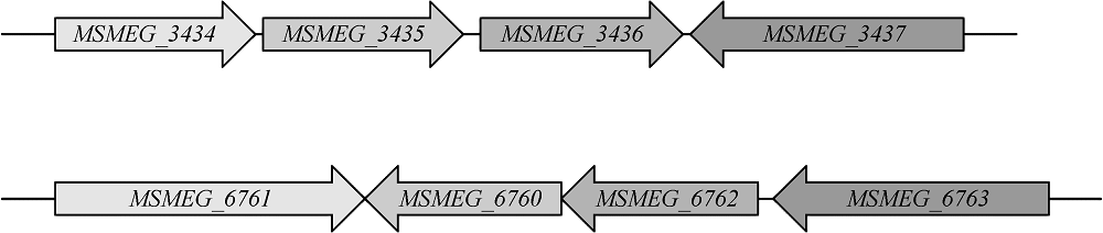
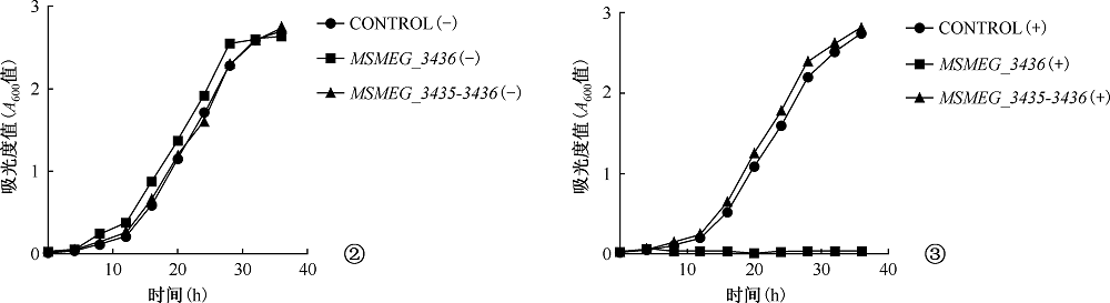
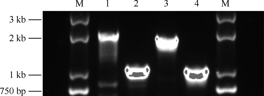
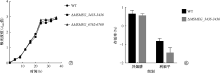
目的 初步探讨耻垢分枝杆菌(Mycobacterium smegmatis)毒素-抗毒素(toxin-antitoxin,TA)系统基因的功能及其在细菌药物耐受中的作用。方法 利用无水四环素(ATc)诱导穿梭质粒构建毒素基因(MSMEG_3436MSMEG_6760)表达系统,检测毒素基因表达的抑菌作用。应用CRISPR-Cas12a基因编辑技术构建ΔMSMEG_3435-3436和ΔMSMEG_6762-6760敲除菌株,探究毒素-抗毒素系统对菌株生长的影响。通过计算菌株存活率检测MSMEG_3435-3436基因对异烟肼(96μg/ml)和利福平(40μg/ml)的耐受相关性。在耻垢分枝杆菌中用LacZ报告基因分别替换毒素-抗毒素基因(MSMEG_1277-1278MSMEG_1283-1284MSMEG_3435-3436MSMEG_4447-4448MSMEG_5635-5634),构建5个启动子活性检测突变菌株(SY3328、SY3309、SY6407、SY3310和SY3311),并将pMV261空载体和pMV261-抗毒素系列质粒分别电转至5个突变菌株中,通过测定吸光度值(A600A550A420)计算β-半乳糖苷酶活性[酶活性单位为“Miller单位(MU)”],以检测毒素抗毒素系统的启动子活性。结果 在耻垢分枝杆菌中,ATc诱导表达毒素基因MSMEG_3436可抑制细菌生长,而同时表达对应的抗毒素基因MSMEG_3435可消除抑制作用;ATc诱导表达毒素基因MSMEG_6760未发现明显的抑菌作用。与野生株相比,ΔMSMEG_3435-3436和ΔMSMEG_6762-6760敲除菌株在7H9液体培养基中生长表型无明显差异。野生株和ΔMSMEG_3435-3436敲除菌株经异烟肼和利福平处理后的存活率[分别为(4.38±1.48)%和(3.49±0.66)%,(0.15±0.04)%和(0.03±0.02)%]显示毒素-抗毒素基因MSMEG_3435-3436与药物耐受性无关(t=0.548,P=0.613;t=2.663,P=0.056)。启动子(SY3328、SY3309、SY6407、SY3310和SY3311)在携带pMV261-空载体和pMV261抗毒素表达质粒的报告菌株中的β-半乳糖苷酶活性分别为(376.50±17.13)和(315.50±20.71)、(189.00±12.24)和(160.70±9.89)、(225.20±9.95)和(211.70±2.57)、(221.40±12.07)和(186.60±13.17)、(179.10±5.87)和(127.70±19.21)MU,差异均无统计学意义(t=2.272,P=0.086;t=1.795,P=0.147;t=1.319,P=0.258;t=1.949,P=0.123;t=2.562,P=0.063)。结论 成功构建了MSMEG_3435-3436MSMEG_6762-6760在耻垢分枝杆菌中的诱导表达体系及敲除菌株,并发现MSMEG_3435-3436是一个新的有功能的毒素-抗毒素系统,以及这两个毒素-抗毒素系统与菌株生长表型及异烟肼和利福平耐受性无关,最后发现耻垢分枝杆菌中5对毒素-抗毒素系统的抗毒素基因可能在自身启动子调控中不发挥关键作用,可为进一步研究结核分枝杆菌毒素-抗毒素系统的功能提供线索。

关键词: 分枝杆菌,耻垢, 类毒素类, 抗毒素类, 基因表达调控, 细菌, 药物耐受性

Abstract:

Objective To investigate the function of the toxin-antitoxin (TA) systems in Mycobacterium smegmatis (M. smegmatis) and its role in bacterial drug tolerance. Methods Two toxin genes (MSMEG_3436 and MSMEG_6760) were constructed in anhydrous tetracycline (ATc)-induced shuttle plasmid, respectively, and then tested for repression of cell growth in M.smegmatis. The ΔMSMEG_3435-3436 and ΔMSMEG_6762-6760 mutants were constructed using CRISPR-Cas12a genome editing system to investigate the effect of TA system on cell growth. Wild type M.smegmatis and MSMEG_3435-3436 deletion mutant were treated with isoniazid (96 μg/ml) and rifampicin (40 μg/ml) and tested for drug tolerance by calculating survival rate. The TA genes (MSMEG_1277-1278, MSMEG_1283-1284, MSMEG_3435-3436, MSMEG_4447-4448, MSMEG_5635-5634) was replaced by LacZ reporter gene in M.smegmatis, resulting in promoter activity analysis stains SY3328, SY3309, SY6407, SY3310, and SY3311. The empty vector pMV261 and its derivatives expressing antitoxins were transformed into the aforementioned strains, respectively. The promoter activity of TA genes was then assessed by measurement of β-galactosidase. Results Highly expression of toxin gene MSMEG_3436 but not MSMEG_6760 in M.smegmatis repressed cell growth, while co-expression of anti-toxin gene MSMEG_3435 relieved the repression. The wild-type, ΔMSMEG_3435-3436 and ΔMSMEG_6762-6760 mutants showed similar growth phenotype in 7H9 liquid medium. Wild-type strain and ΔMSMEG_3435-3436 mutant treated with isoniazid and rifampicin had similar survival rate ((4.38±1.48) % and (3.49±0.66) %, (0.15±0.04) % and (0.03±0.02) %), which suggested that MSMEG_3435-3436 might not play an important role in drug tolerance of M.smegmatis (t=0.548, P=0.613; t=2.663, P=0.056). Promoter activity analysis showed that the β-galactosidase activities ((376.50±17.13) and (315.50±20.71) Miller units (MU), (189.00±12.24) and (160.70±9.89) MU, (225.20±9.95) and (211.70±2.57) MU, (221.40±12.07) and (186.60±13.17) MU, (179.10±5.87) and (127.70±19.21) MU) in the reporter strains harboring empty vector and antitoxin expressing plasmids were not significantly different (t=2.272, P=0.086; t=1.795, P=0.147; t=1.319, P=0.258; t=1.949, P=0.123; t=2.562, P=0.063). Conclusion The MSMEG_3435-3436 and MSMEG_6762-6760 induced expression systems and knockout strains are constructed in M.smegmatis. Further study shows that MSMEG_3435-3436 is a functional toxin-antitoxin system, but does not affect cell growth and might not affect the tolerance to isoniazid and rifampicin in M.smegmatis. Finally, the antitoxins of the known 5 TA systems are found not critical for auto-regulation of their promoter’s activity in M.smegmatis. These findings provide clues for further investigation of the roles of TA systems in M.tuberculosis.

Key words: Mycobacterium smegmatis, Toxoids, Antitoxins, Gene expression regulation, bacterial, Drug tolerance