Email Alert | RSS    帮助

中国防痨杂志 ›› 2020, Vol. 42 ›› Issue (1): 31-37.doi: 10.3969/j.issn.1000-6621.2020.01.009

• 论著 • 上一篇    下一篇

艾滋病并发颈部淋巴结结核的临床特征及CT表现

卢亦波(),周静如,莫移美,宋树林   

  1. 530023 南宁市第四人民医院 西艾滋病临床治疗中心放射科
  • 收稿日期:2019-12-02 出版日期:2020-01-10 发布日期:2020-01-08
  • 通信作者: 卢亦波 E-mail:13768415203@139.com
  • 基金资助:
    南宁市兴宁区科学研究与技术开发项目(2018A06);南宁市科学研究与技术开发计划基金(20173157-6)

Clinical features and manifestations of AIDS complicated with cervical lymph node tuberculosis

LU Yi-bo(),ZHOU Jing-ru,MO Yi-mei,SONG Shu-lin   

  1. Department of Radiology,the Fourth People’s Hospital of Nanning(AIDS Clinical Treatment Center of Guangxi),Nanning 530023,China
  • Received:2019-12-02 Online:2020-01-10 Published:2020-01-08
  • Contact: Yi-bo LU E-mail:13768415203@139.com

摘要:

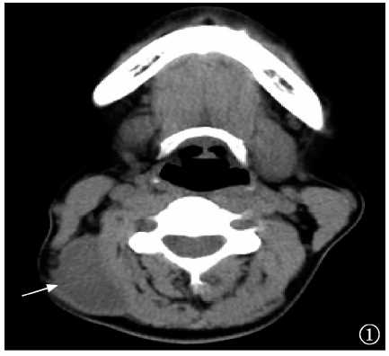
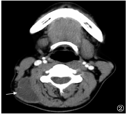
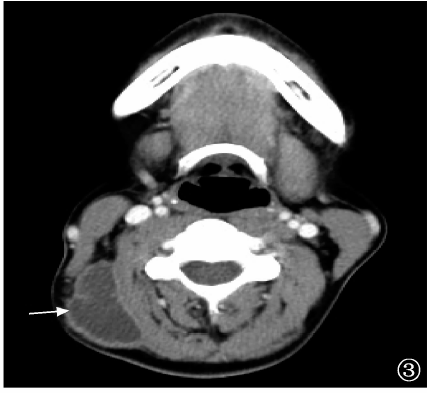
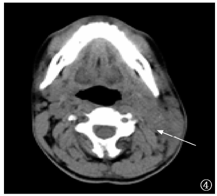
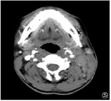
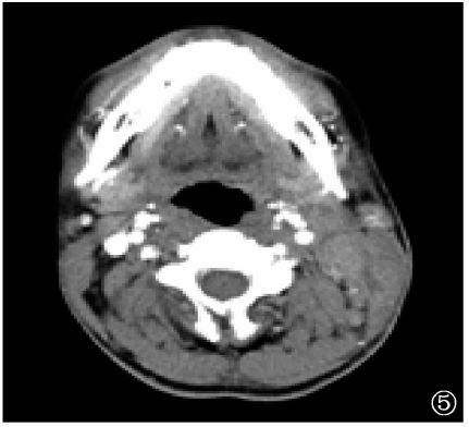
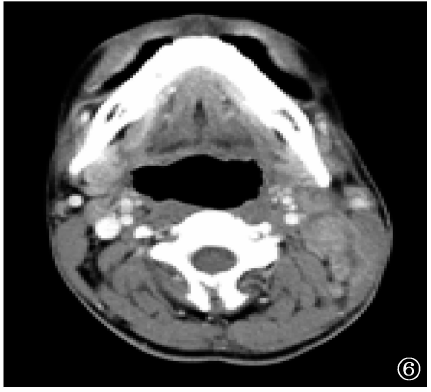
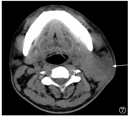
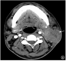
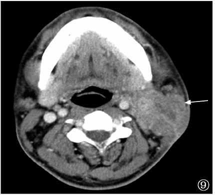
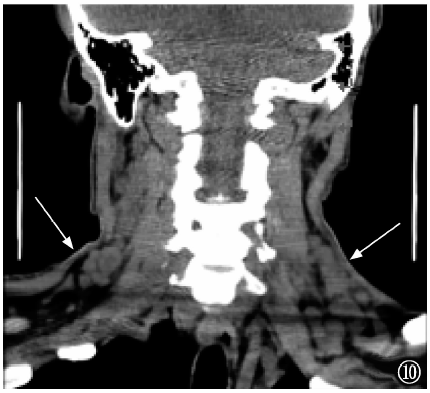
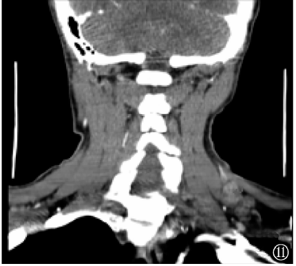
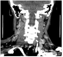
目的 探讨艾滋病(AIDS)并发颈部淋巴结结核的临床特征及CT表现。方法 收集2015年1月至2019年7月南宁市第四人民医院经颈部淋巴结穿刺活检或手术后组织病理检查确诊为颈部淋巴结结核的142例临床资料完整的初治住院患者,按照是否并发AIDS分为并发组(AIDS并发颈部淋巴结结核,42例)和未并发组(未并发AIDS的颈部淋巴结结核,100例),对比分析两组患者的临床特征及CT表现。结果 并发组患者在男性[66.7%(28/42]、年龄[41.69(34.75,47.00)岁]、咳嗽[26.2%(11/42)]、乏力[16.7%(7/42)]、发热[38.1%(16/42)]等方面均明显高于未并发组[分别为44.0%(44/100)、30.64(22.00,32.75)岁、9.0%(9/100)、1.0%(1/100)、8.0%(8/100)](χ 2=6.867,P=0.009;Z=-5.300,P=0.000;χ 2=7.223,P=0.007;χ 2=10.867,P=0.001;χ 2=13.339,P=0.000),而CD4 + T淋巴细胞计数[152.50(69.25,241.75)个/μl]明显低于未并发组[598.00(452.00,748.00)个/μl](Z=-8.081,P=0.001)。在CT扫描表现上,并发组在淋巴结短径>2cm[47.6%(20/42)]、淋巴结融合[71.4%(30/42)]、淋巴结完全性与不完全性坏死并存型[45.2%(19/42)]、累及≥3个区[59.5%(25/42)]、累及≥4个区[33.3%(14/42)]等方面均明显高于未并发组[27.0%(27/100)、53.0%(53/100)、4.0%(4/100)、31.0%(31/100)、11.0%11/100)](χ 2值分别为5.679、4.136、37.056、10.075、10.170,P值均<0.05);在形态规则[16.7%(7/42)]、边界清楚[16.7%(7/42)]、不全性坏死[33.3%(14/42)]方面均明显低于未并发组[50.0%(50/100)、52.0%(52/100)、90.0%(90/100)](χ 2值分别为13.677、15.205、48.459,P值均=0.000)。结论 AIDS并发颈部淋巴结结核以男性、40岁以上患者多见;临床表现不典型,CD4 + T淋巴细胞计数明显降低。CT表现多见淋巴结短径>2cm、融合、边界不清、形态不规则,病变常累及≥3个分区的淋巴结,且多为并存型坏死。

关键词: 获得性免疫缺陷综合征, 结核,淋巴结, 共病现象, 体层摄影术,X线计算机, 疾病特征

Abstract:

Objective To investigate the clinical features and CT characteristics of acquired immune deficiency syndrome (AIDS) complicated with cervical lymph node tuberculosis. Methods From Jan 2015 to July 2019, 142 clinical data of initial inpatients who diagnosed with cervical lymph node tuberculosis by cervical lymph node biopsy or postoperative histopathological examination at the Fourth People’ Hospital of Nanning were collected. These patients were divided into two groups based on whether complicated with AIDS: complicated group (AIDS with cervical lymph node tuberculosis, 42 cases) and non-complicated group (AIDS without cervical cancer without tuberculosis, 100 cases), and then the clinical characteristics and CT manifestation of the two groups were compared and analyzed. Results In the complicated group, several factors, including the proportion of male, age, cough ratio, fatigue ratio and fever ratio, were significantly higher than those in the non-complicated group (66.7% (28/42) vs 44.0% (44/100), χ 2=6.867, P=0.009; 41.69 (34.75, 47.00) years vs 30.64 (22.00, 32.75) years, Z=-5.300, P=0.000; 26.2% (11/42) vs 9.0% (9/100), χ 2=7.223, P=0.007; 16.7% (7/42) vs 1.0% (1/100), χ 2=10.867, P=0.001; and 38.1% (16/42) vs 8.0% (8/100), χ 2=13.339, P=0.000, respectively); however, the CD4 + T lymphocyte count (152.50 (69.25, 241.75) cells/μl) in the complicated group was obviously lower than that (598.00 (452.00, 748.00) cells/μl) in the non-complicated group (Z=-8.081, P=0.001). In addition, in the complicated group, several CT manifestations, including lymph nodes with a short diameter >2 cm (47.6% (20/42)), lymph node fusion (71.4% (30/42)), coexistence of complete and incomplete necrosis of the lymph nodes (45.2% (19/42)), involvement ≥3 areas (59.5% (25/42)) and involvement ≥4 areas (33.3% (14/42)), were significantly higher than those in the non-complicated group (27.0% (27/100), 53.0% (53/100), 4.0% (4/100), 31.0% (31/100), and 11.0% (11/100), respectively) (χ 2= 5.679, 4.136, 37.056, 10.075, and 10.170, respectively, all P<0.05), while the regular lesions (16.7% (7/42)), the clear borders (16.7% (7/42)), and incomplete necrosis (33.3% (14/42)) were remarkably lower in the complicated group than those (50.0% (50/100), 52.0% (52/100), and 90.0% (90/100)) in the non-complicated group (χ 2=13.677, 15.205, and 48.459, respectively, all P=0.000). Conclusion AIDS complicated with cervical lymph node tuberculosis usually occurred in males and patients over 40 years of age, as well as exhibited atypical clinical characteristics and significantly reduced CD4 + T lymphocyte counts. CT manifestations of these patients usually exhibited are more common for lymph nodes with a short diameter >2 cm, fusion, unclear borders, irregular morphology, lesions involved lymph nodes ≥ 3 areas, and more mixed necrosis.

Key words: Acquired immunodeficiency syndrome, Tuberculosis,lymph node, Comorbidity, Tomogrphy,X-ray computer, Disease attributes