Email Alert | RSS    帮助

中国防痨杂志 ›› 2019, Vol. 41 ›› Issue (7): 715-718.doi: 10.3969/j.issn.1000-6621.2019.07.003

• 论著 • 上一篇    下一篇

超声造影在疑似胸壁结核患者穿刺活检术中的应用价值

张文智(),苏冬明,孟君,何宁,王彩芬   

  1. 310003 杭州,浙江省中西医结合医院超声科
  • 收稿日期:2019-06-18 出版日期:2019-07-10 发布日期:2019-07-09
  • 通信作者: 张文智 E-mail:zwz2009999@163.com
  • 基金资助:
    杭州市社会发展自主申报项目(20170533B71);杭州市农业与社会发展科研主动设计项目(20190101A09);杭州市科技计划引导项目(20163501Y33)

Application and value of contrast-enhanced ultrasound in puncture biopsy of patients with chest wall tuberculosis

Wen-zhi ZHANG(),Dong-ming SU,Jun MENG,Ning HE,Cai-fen WANG   

  1. Department of Ultrasound, Integrated Chinese and Western Medicine Hospital of Zhejiang Province, Hangzhou 310003, China
  • Received:2019-06-18 Online:2019-07-10 Published:2019-07-09
  • Contact: Wen-zhi ZHANG E-mail:zwz2009999@163.com

摘要:

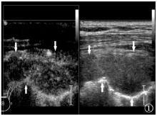
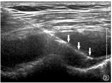
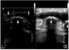
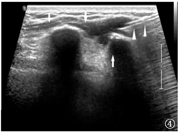
目的 探讨超声造影在疑似胸壁结核患者穿刺活检术中的应用及其临床价值。方法 选取2017年4月至2019年6月入住浙江省中西医结合医院的疑似胸壁结核(均局部形成肿块)患者78例,男31例,女47例;年龄18~67岁,平均年龄(33.8±4.7)岁。依据时间对照法,将患者分为两组:A组42例,2017年4月至2018年5月入住我院的疑似胸壁结核患者,依据超声扫描所示行超声引导下组织学穿刺活检术,并对无回声区行穿刺抽液术;B组36例,2018年1月至2019年6月入住我院的疑似胸壁结核患者,先行超声造影,依据超声造影表现在超声选择性引导下行穿刺抽液术或组织学穿刺活检术。对所有患者的超声表现及病理结果进行回顾性分析。两组患者获取的组织标本送病理检查,同时送MTB、普通细菌培养及GeneXpert MTB/RIF检测。结果 A组:42例患者穿刺取出脓液的成功率为77.4%(24/31),获取活检组织标本的成功率为88.1%(37/42)。B组:36例患者穿刺取出脓液的成功率为100.0%(23/23),获取活检组织标本的成功率为100.0%(36/36),较A组均明显提高(χ 2值分别为4.132和2.814,P值分别为0.001和0.003)。A、B两组患者诊断为胸壁结核在内所有疾病的诊断阳性率分别为83.3%(35/42,其中确诊为胸壁结核33例)和100.0%(36/36,其中确诊为胸壁结核34例),B组较A组诊断阳性率明显提高(χ 2=4.713,P=0.001)。结论 超声造影在疑似胸壁结核患者穿刺活检术中的临床应用价值突出,对提高诊断阳性率有重要价值。

关键词: 结核,胸膜, 胸壁, 超声检查,介入性, 活组织检查,针吸, 对比研究, 超声造影

Abstract:

Objective To investigate the application and clinical value of contrast-enhanced ultrasound in puncture biopsy of patients with chest wall tuberculosis (TB). Methods Between April 2017 and June 2019, 78 patients with suspected TB of chest wall were admitted to Integrated Chinese and Western Medicine Hospital of Zhejiang Province,among them 31 were male patients, 47 were female cases, ages ranged from 18 to 67 years, mean age (33.8±4.7) years. According to the time control method, patients were divided into two groups: Group A of 42 cases, the suspected chest wall TB patients admitted between April 2017 and May 2018 to our hospital, on the basis of the results of ultrasound scanning, ultrasound-guided histological puncture biopsy was performed and line puncture pumping fluid were performed in the anechoic area; Group B of 36 cases, the suspected chest wall TB patients admitted between January 2018 and June 2019 to our hospital. Contrast-enhanced ultrasonography (CEUS) was performed first, and ultrasound-guided piercing or histological biopsy was performed according to the findings of CEUS. The ultrasound manifestations and pathological results of all patients were retrospectively analyzed. Tissue samples of the two groups of patients were sent for pathologic examination, at the same time for MTB, ordinary culture and GeneXpert MTB/RIF examination. Results Group A: the success rate with piercing out pus was 77.4% (24/31) in 42 cases, the success rate for obtaining biopsy tissue samples was 88.1% (37/42);Group B: the success rate with piercing out pus was 100.0% (23/23) in 36 cases, the success rate for obtaining biopsy tissue samples was 100.0% (36/36). Group B was obviously improved (χ 2 values were 4.132 and 2.814, respectively, P values were 0.001 and 0.003). The positive rate of two groups of patients with all disease diagnosis was 83.3% (35/42, 33 cases diagnosed as chest wall TB) and 100.0% (36/36, 34 cases diagnosed as chest wall TB) respectively, group B was significantly higher than group A (χ 2=4.713, P=0.001). Conclusion The clinical application value of contrast-enhanced ultrasound in biopsy patients with suspected chest wall TB is important to improve the diagnostic positive rate.

Key words: Tuberculosis,pleural, Thoracic wall, Ultrasonography,interventional, Biopsy,needle, Comparative study, Contrast-enhanced ultrasound