Email Alert | RSS    帮助

中国防痨杂志 ›› 2019, Vol. 41 ›› Issue (2): 202-209.doi: 10.3969/j.issn.1000-6621.2019.02.015

• 论著 • 上一篇    下一篇

非结核分枝杆菌肺病的CT分型及其临床特征分析(附132例报告)

李多,房坤,王珏,周震,吕平欣()   

  1. 100149 首都医科大学附属北京胸科医院影像科
  • 收稿日期:2018-07-01 出版日期:2019-02-10 发布日期:2019-02-01
  • 通信作者: 吕平欣 E-mail:lpx1209@163.com
  • 基金资助:
    北京市优秀人才项目(2017000021469G283)

Analysis of CT image classification and clinical characteristics of nontuberculous mycobacterial pulmonary disease

Duo LI,Kun FANG,Jue WANG,Zhen ZHOU,Ping-xin LYU()   

  1. Department of Radiology,Beijing Chest Hospital, Capital Medical University, Beijing 101149, China
  • Received:2018-07-01 Online:2019-02-10 Published:2019-02-01
  • Contact: Ping-xin LYU E-mail:lpx1209@163.com

摘要:

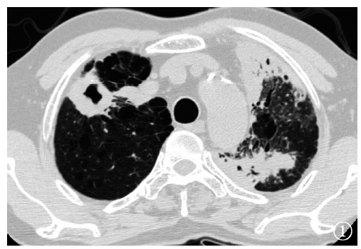
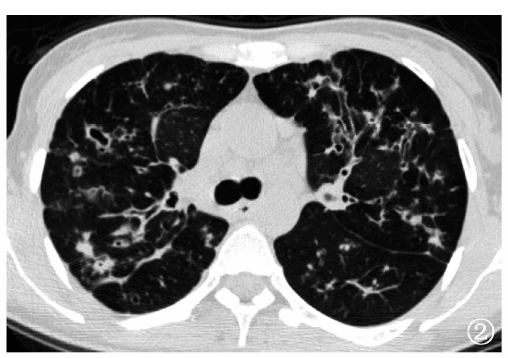
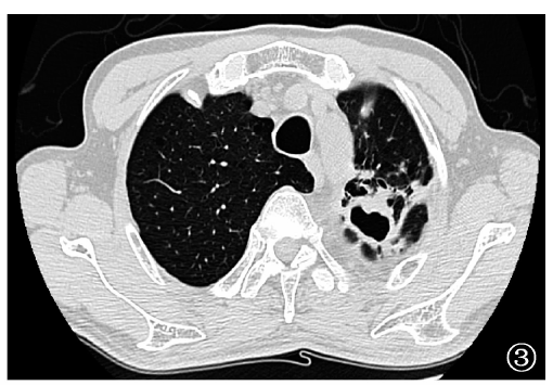
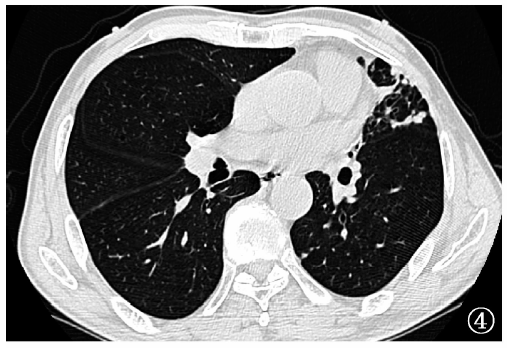
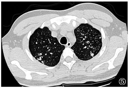
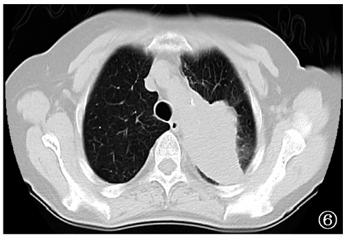
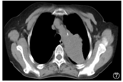
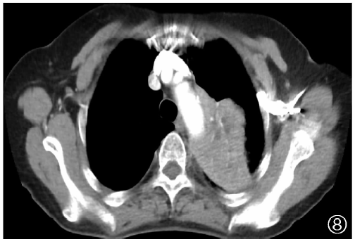
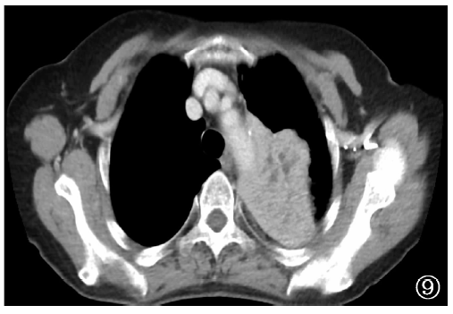
目的 分析非结核分枝杆菌肺病的CT分型,以及不同型别患者的临床及CT表现特点。方法 回顾性分析2011年11月至2018年1月首都医科大学附属北京胸科医院确诊的132例非结核分枝杆菌肺病患者的临床及CT资料,根据CT表现分为上叶空洞型、结节支气管扩张型、混合型和不易分类型4种类型,统计分析不同CT分型患者的临床及CT特征。采用SPSS 17.0软件进行统计学分析,计数资料采用χ 2检验或连续校正χ 2检验,计量资料采用t检验或方差分析,均以P<0.05为差异有统计学意义;4组不同CT型别患者的临床统计数据进行两两比较时,以P<0.008为差异有统计学意义。结果 132例非结核分枝杆菌肺病CT分型结果为:上叶空洞型36例(27.3%),结节支气管扩张型61例(46.2%),混合型15例(11.4%),不易分类型20例(15.2%)。非结核分枝杆菌肺病不同CT分型组中,男性构成比由高到低分别为上叶空洞型(86.1%,31/36)、不易分类型(70.0%,14/20),混合型(53.3%,8/15)和结节支气管扩张型(24.6%,15/61),差异有统计学意义(χ 2=37.712,P<0.001)。上叶空洞型、结节支气管扩张型、混合型和不易分类型患者平均年龄分别为(58.5±13.8)岁、(58.1±13.3)岁、(61.3±10.4)岁和(51.0±17.0)岁,差异无统计学意义(F=1.875,P=0.140)。上叶空洞型患者中吸烟者占64.7%(22/34),明显高于结节支气管扩张型患者(13.0%,7/54)(χ 2=25.258,P<0.001)。上叶空洞型患者发生肺气肿者占66.7%(24/36),发生间质纤维化改变者占33.3%(12/36),明显高于结节支气管扩张型[分别为13.1%(8/61)和0.0%(0/61)](χ 2=29.369、23.204,P值均<0.001)。上叶空洞型并发胸膜增厚者占61.1%(22/36),明显高于结节支气管扩张型(23.0%,14/61)(χ 2=14.125,P<0.001);混合型患者并发胸膜增厚者占66.7%(10/15),同样高于结节支气管扩张型(χ 2=10.649,P=0.001)。结论 非结核分枝杆菌肺病中,结节支气管扩张型最常见;上叶空洞型好发于男性有肺气肿和(或)间质纤维化改变的患者,结节支气管扩张型好发于女性无肺部基础疾病(未并发肺部其他种类疾病,包括肺气肿、间质纤维化等)的患者。

关键词: 分枝杆菌感染, 非典型性, 肺疾病, 体层摄影术, X线计算机, 诊断显像

Abstract:

Objective To analyze the CT image classification of non-tuberculosis mycobacterium(NTM) pulmonary disease and the clinical and CT features in patients with different CT image classifications. Methods The clinical and CT data of 132 patients with non-tuberculous mycobacterial pulmonary disease admitted in Beijing Chest Hospital, Capital Medical University from November 2011 to January 2018 were analyzed retrospectively. According to CT imaging features, there were four types of cavities in the upper lobe, nodular bronchiectasis, mixed and difficult to differentiate. The clinical and imaging characteristics of patients with different image classification were analyzed statistically using SPSS software with 17.0 version, and the enumeration data were analyzed by chi-square test or continuous correction chi-square test. Measurement data were analyzed by t test or variance analysis. The difference were considered statistically significant when P<0.05. When the clinical statistical data of 4 groups of patients with different types of patients were compared in pairs, P<0.008 was considered as the significant statistically difference. Results Of 132 cases with non-tuberculous mycobacterial pulmonary disease, there were 36 (27.3%) cases in the type of upper lobe cavitary 61 (46.2%) cases in the type of nodular bronchiectasis, 15 (11.4%) cases in the mixed type and 20 (15.2%) cases in unclassified type. The male ratio from high to low were upper lobe cavitary (86.1%, 31/36), unclassified type (70.0%, 14/20), mixed type (53.3%, 8/15) and nodular bronchiectasis (24.6%, 15/61) in all cases with significant statistically difference (χ 2=37.712,P<0.001).The average age of patients with upper lobe cavitary, nodular bronchiectasis, mixed type and unclassified type were 58.5±13.8, 58.1±13.3, 64.3±10.4 and 51.0±17.0 years old with no significant difference (F=1.875, P=0.140). The ratio of smoker in patients with upper lobe cavitary (64.7%,22/34) was significantly higher than that in patients with nodular bronchiectatic type (13%, 7/54)(χ 2=25.258, P<0.001). The incidence rate of emphysema (66.7%,24/36) and interstitial fibrosis (33.3%,12/36) in patients with upper lobe cavitary were significant higher than those (13.1%,8/61 and 0.0%,0/61) in patients with nodular bronchiectasis (χ 2=29.369,P<0.001;χ 2=23.204,P<0.001). The incidence rates of pleural thickening in upper lobe cavity type (61.1%, 22/36) and in mixed type (66.7%,10/15) were higher than that in nodular bronchiectasis (23.0%,14/61) (χ 2=10.649,P=0.001). Conclusion Nodular bronchiectasis type is the most common imaging features in NTM pulmonary disease. The upper lobe cavitary type is more common in male patients with emphysema and/or interstitial fibrosis. Nodular bronchiectatic type is frequently seen in female patients without underlying lung diseases.

Key words: Mycobacterium infections, atypical, Lung diseases, Tomography, X-ray computed, Diagnostic imaging