Email Alert | RSS    帮助

中国防痨杂志 ›› 2019, Vol. 41 ›› Issue (1): 57-63.doi: 10.3969/j.issn.1000-6621.2019.01.013

• 论著 • 上一篇    下一篇

伴空洞的胞内分枝杆菌肺病与继发性肺结核的CT表现对比分析

杨佳,吕圣秀(),李春华,舒伟强,王惠秋,唐光孝,刘雪艳   

  1. 400036 重庆市公共卫生医疗救治中心放射科
  • 收稿日期:2018-07-05 出版日期:2019-01-10 发布日期:2019-01-09

Comparative analysis of CT findings between Mycobacteria intracellulare lung disease and pulmonary tuberculosis both with cavitation

Jia YANG,Sheng-xiu LYU(),Chun-hua LI,Wei-qiang SHU,Hui-qiu WANG,Guang-xiao TANG,Xue-yan LIU   

  1. Department of Radiology, Chongqing Public Health Medical Center, Chongqing 400036, China
  • Received:2018-07-05 Online:2019-01-10 Published:2019-01-09

摘要:

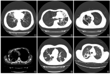
目的 探讨伴空洞的胞内分枝杆菌肺病与继发性肺结核的CT表现差异。方法 搜集2016年6月至2018年3月重庆市公共卫生医疗救治中心经临床及实验室检查确诊,符合纳入标准(具有治疗前完整临床及影像学资料,既往未经过抗NTM及抗结核药物治疗,排除并发尘肺、糖尿病、HIV或其他感染,且均伴有直径>10mm空洞者)的全部胞内分枝杆菌肺病患者26例作为观察组;采用随机数字表法在同期符合纳入标准(纳入标准与观察组相同)的862例继发性肺结核患者中抽取40例患者作为对照组。对两组患者CT检查表现的支气管扩张分类及分布、空洞形态及邻近胸膜增厚、肺体积缩小、肺气肿、纵隔淋巴结肿大等情况进行统计学分析。结果 观察组发生支气管扩张、静脉曲张状及囊状支气管扩张、肺部病灶钙化、肺体积缩小、肺气肿、薄壁空洞、空洞邻近胸膜增厚分别占92.3%(24/26)、88.5%(23/26)、57.7%(15/26)、69.2%(18/26)、57.7%(15/26)、73.1%(19/26)、80.8%(21/26),均明显多于对照组[分别占60.0%(24/40)、35.0%(14/40)、15.0%(6/40)、15.0%(6/40)、10.0%(4/40)、25.0%(10/40)、37.5%(15/40)],差异均有统计学意义(χ 2值分别为8.29、18.28、13.24、20.03、17.48、14.79、11.90,P值均<0.05);观察组大结节影(直径≥10mm)、结节边缘模糊、单发空洞、厚壁空洞、纵隔淋巴结肿大、心包积液及心包增厚者分别占19.2%(5/26)、34.6%(9/26)、7.7%(2/26)、26.9%(7/26)、23.1%(6/26)、7.7%(2/26),均明显少于对照组[分别占57.5%(23/40)、72.5%(29/40)、37.5%(15/40)、75.0%(30/40)、47.5%(19/40)、30.0%(12/40)],差异均有统计学意义(χ 2值分别为9.45、9.26、7.32、14.79、3.99、4.69,P值均<0.05)。观察组无支气管扩张、支气管扩张占1~2叶的发生率分别为11.5%(3/26)、19.2%(5/26),均明显低于对照组[分别为40.0%(16/40)、50.0%(20/40)],差异均有统计学意义(χ 2值分别为6.23、6.34,P值均<0.05);观察组支气管扩张占3~4叶、占≥5叶的发生率分别为30.8%(8/26)、38.5%(10/26),均明显高于对照组[分别为5.0%(2/40)、5.0%(2/40)],差异均有统计学意义(连续校正χ 2值分别为6.26、9.72,P值均<0.05);观察组静脉曲张状及囊状支气管扩张占3~4叶的发生率为26.9%(7/26),明显高于对照组(5.0%,2/40),差异有统计学意义(连续校正χ 2值为4.70,P<0.05)。 结论 伴有空洞的胞内分枝杆菌肺病患者CT表现中薄壁空洞、肺体积缩小、肺气肿、双肺广泛静脉曲张状及囊状支气管扩张多于继发性肺结核患者,大结节影(直径>10mm)、结节边缘模糊、单发空洞、厚壁空洞、纵隔淋巴结肿大、心包积液少于继发性肺结核患者,以上特征有助于两种疾病的鉴别诊断。

关键词: 分枝杆菌感染, 非典型性, 结核, 肺, 体层摄影术, X线计算机, 诊断显像, 对比研究

Abstract:

Objective To discuss the difference in CT manifestations of Mycobacteria intracellulare lung di-sease and pulmonary tuberculosis both with cavitation.Methods Twenty-six cases of Mycobacteria intracellulare lung disease(Observation group) and 40 cases of pulmonary tuberculosis (Control group) accompanied by a cavitation of above 10 mm were studied. Forty patients with pulmonary tuberculosis included in the study were chosen randomly from 862 cases of secondary pulmonary tuberculosis, which were diagnosed with clinical examinations and lab tests in the Chongqing Public Health Medical Center from June 2016 to March 2018. The cases all had complete clinical and imaging data before treatment, and did not receive anti-NTM and tuberculosis treatments. Cases combined with pneumoconiosis, diabetes, HIV or other infections were excluded. Statistical analysis was conducted on CT findings of the two groups, including classification and distribution of bronchiectasis, cavitation morphology and neighboring pleural thickening, lung volume reduction,emphysema and mediastinal lymph node enlargement.Results In the observation group, bronchiectasis (92.3%, 24/26) including varicose bronchiectasis and cystic bronchiectasis (88.5%, 23/26), calcification of lung lesions (57.7%, 15/26), lung volume reduction (69.2%, 18/26), emphysema (57.7%, 15/26), thin-walled cavities (73.1%, 19/26), and pleural thickening adjacent to the cavities (80.8%, 21/26) were all more than those in the control group (60.0% (24/40), 35.0% (14/40), 15.0% (6/40), 15.0% (6/40), 10.0% (4/40), 25.0% (10/40) and 37.5% (15/40)), which were statistically significant (χ 2=8.29, 18.28, 13.24, 20.03, 17.48, 14.79 and 11.90 respectively, P<0.05); in the observation group, large nodules (diameter ≥10 mm) 19.2% (5/26), infiltrates around the nodules (34.6%, 9/26), single cavities (7.7%, 2/26), thick-walled cavities (26.9%, 7/26), mediastinal lymph node enlargement (23.1%, 6/26) and pericardial effusion (7.7%, 2/26) were all significantly lower than those in the control group (57.5% (23/40), 72.5% (29/40), 37.5% (15/40), 75.0% (30/40), 47.5% (19/40), and 30.0% (12/40)) (χ 2=9.45,9.26,7.32,14.79,3.99 and 4.69 respectively, P<0.05). In the observation group, the incidences of 0-lobe and 1-2-lobe bronchiectasis 11.5% (3/26) and 19.2% (5/26) respectively were lower than those in the control group 40.0% (16/40) and 50.0% (20/40) respectively, the differences were statistically significant (χ 2=6.23 and 6.34, P<0.05); the incidence of 3-4-lobe and ≥5-lobe bronchiectasis 30.8% (8/26) and 38.5% (10/26) respectively were higher than those in the control group (5.0% (2/40) and 5.0% (2/40) respectively), the differences were statistically significant (continuity correction χ 2=6.26 and 9.72, respectively, P<0.05); In the observation group, the incidence of 3-4-lobe bronchiectasis which were varicose and cystic types 26.9% (7/26) was higher than those in the control group 5.0% (2/40) (continuity correction, χ 2=4.70, P<0.05). Conclusion The thin-walled cavities, lung volume reduction, emphysema, extensive varicose bronchiectasis and cystic bronchiectasis of Mycobacteria intracellulare lung disease with cavitation are all more than pulmonary tuberculosis, large nodules (diameter ≥10 mm), infiltrates around the nodules, single cavities, thick-walled cavities, mediastinal lymph node enlargement and pericardial effusion are all significantly lower than pulmonary tuberculosis in the CT manifestations, which is helpful for differentiation between the two groups.

Key words: Mycobacterium infections, atypical, Tuberculosis, pulmonary, Tomography, X-ray, Diagnostic imaging, Comparative study computed