Email Alert | RSS    帮助

中国防痨杂志 ›› 2018, Vol. 40 ›› Issue (7): 677-681.doi: 10.3969/j.issn.1000-6621.2018.07.003

• 论著 • 上一篇    下一篇

结核性胸膜炎发病早期的CT征象及其动态变化特点的分析

王东坡,杨新婷,吕岩,王珏,房坤,周新华,陈步东()   

  1. 101149 首都医科大学附属北京胸科医院放射科(王东坡、吕岩、王珏、房坤、周新华、陈步东),结核三科(杨新婷);
  • 收稿日期:2018-03-20 出版日期:2018-07-10 发布日期:2018-09-07
  • 通信作者: 陈步东 E-mail:budongchen@sina.com
  • 基金资助:
    北京市科学技术委员会重点项目(D14117005214003)

Analysis of CT signs and dynamic changes of tuberculous pleurisy at the early stage of onset

Dong-po WANG,Xin-ting YANG,Yan LYU,Jue WANG,Kun FANG,Xin-hua ZHOU,Bu-dong CHEN()   

  1. Department of Radiology, Beijing Chest Hospital, Capital Medicine University, Beijing 101149, China
  • Received:2018-03-20 Online:2018-07-10 Published:2018-09-07
  • Contact: Bu-dong CHEN E-mail:budongchen@sina.com

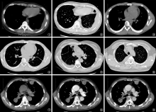
摘要:

目的 分析结核性胸膜炎发病早期的CT征象及其动态演变的特点,提高诊断准确率。方法 收集首都医科大学附属北京胸科医院于2015年3月至2017年2月确诊为结核性胸膜炎的38例患者的影像资料。研究对象中包括男16例,女22例;年龄21~61岁,平均年龄为(35.74±11.92)岁;所有患者均在初次发病7~10d内就诊。分析研究对象发病早期的CT检查资料,提取CT表现特点及征象。结果 38例研究对象中,发生于单侧胸膜病变者34例。其中,病变位于左侧胸膜者18例,位于右侧胸膜者16例,双侧同时出现病变者4例;累及纵隔胸膜者14例(累及左侧纵隔胸膜者6例,累及右侧纵隔胸膜者8例),累及叶间裂胸膜者34例;出现包裹性胸腔积液者38例,出现胸膜下小叶间隔增厚者24例,出现胸膜下条索状影者24例。CT动态随访过程中,37例患者胸膜增厚程度减轻;1例患者在开始治疗6个月复查时增厚程度加重,最厚约1.1cm,12个月再次复查增厚的胸膜较前吸收减轻。随访过程中所有患者胸腔积液均表现吸收减少,胸膜下小叶间隔增厚表现为吸收减少,胸膜下条索状影亦逐渐减少、减薄。另有22例患者在随访中出现胸膜结核瘤。结论 结核性胸膜炎发病早期行CT检查可发现单侧胸膜增厚且不光滑、叶间裂受累伴多发粟粒状改变及微结节,以及包裹性胸腔积液、胸膜下小叶间隔增厚及条索状影等征象,可作为其诊断依据。

关键词: 结核, 胸膜, 体层摄影术, X线计算机, 疾病特征, 诊断显像

Abstract:

Objective To analyze the characteristics of CT signs and dynamic changes of early-stage tuberculous pleurisy, and to improve the accuracy of diagnosis.Methods The imaging data of 38 patients diagnosed as tuberculous pleurisy in Beijing Chest Hospital affiliated to Capital Medical University from March 2015 to February 2017 were collected. The subjects included 16 men and 22 women. The age was ranged 21-61 years, and the average age was (35.74±11.92) years. All the patients were treated within 7-10 days of the first onset. The CT image data of the early stage of the disease were analyzed, and the imaging features and signs were extracted.Results Among the 38 subjects, there were 34 cases with unilateral pleural lesions (including 18 in the left pleura and 16 in the right pleural) and 4 with bilateral lesions; there were 14 patients involving the mediastinal pleura (including 6 in the left mediastinal pleural and 8 in the right mediastinal pleura), and 34 involving the interlobular pleura. There were 38 cases showing encapsulated pleural effusion, 24 cases showing pleural septal thickening, and 24 cases showing pleural stripe. During the dynamic follow-up period, the thickening of the pleura was reduced in 37 patients. In 1 case, the thickening degree was aggravated at 6 months, and the thickest was about 1.1 cm. The thickening of pleura was rechecked at 12 months, and it was found to become thinned. During follow-up, the pleural effusion of all patients showed decreased absorption, the pleural septal thickening showed decreased absorption, and the pleural stripe was gradually reduced and thinned. Another 22 patients had pleural tuberculoma during follow-up.Conclusion In the early stage of the disease, unilateral pleural thickening and unsmoothness, interlobular fissure involvement, multiple miliary and micronodules, encapsulated pleural effusion, pleural septal thickening and stripe were found in CT images. We may consider tuberculous pleurisy.

Key words: Tuberculosis, pleural, Tomography, X-ray computed, Disease attributes, Diagnostic imaging