Email Alert | RSS    帮助

中国防痨杂志 ›› 2018, Vol. 40 ›› Issue (5): 499-505.doi: 10.3969/j.issn.1000-6621.2018.05.012

• 论著 • 上一篇    下一篇

非结核分枝杆菌肺病和活动性肺结核的高分辨率CT表现异同性分析

李芳,贺伟(),周新华,赵春生,吕岩,李成海,王东坡   

  1. 101149 首都医科大学附属北京胸科医院放射科
  • 收稿日期:2018-03-21 出版日期:2018-05-10 发布日期:2018-06-12
  • 通信作者: 贺伟 E-mail:hewei7734@sina.com

The similarities and differences of HRCT imaging features between nontuberculous mycobacterial lung diseases and active pulmonary tuberculosis

Fang LI,Wei HE(),Xin-hua ZHOU,Chun-sheng ZHAO,Yan LYU,Cheng-hai LI,Dong-po. WANG   

  1. Department of Radiology,Beijing Chest Hospital, Capital Medical University,Beijing 101149,China
  • Received:2018-03-21 Online:2018-05-10 Published:2018-06-12
  • Contact: Wei HE E-mail:hewei7734@sina.com

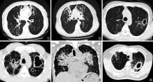
摘要:

目的 探讨非结核分枝杆菌肺病和活动性继发性肺结核的高分辨率CT(HRCT)表现异同性。方法 回顾性分析2012年1月至2017年12月首都医科大学附属北京胸科医院住院并经临床及实验室检查确诊为非结核分枝杆菌肺病患者74例(NTM肺病组)和初治活动性继发性肺结核患者100例(肺结核组)的HRCT表现,比较两组病变分布及HRCT表现。采用SPSS 17.0软件进行统计学分析,计数资料采用χ 2检验,以P<0.05为差异有统计学意义。结果 两组患者CT分型比较,NTM肺病组结节-支气管扩张型(51.4%,38/74)多于肺结核组(14.0%,14/100),两组比较差异有统计学意义(χ 2=28.316, P=0.000);肺结核组结节-肿块型(21.0%,21/100)多于NTM肺病组(8.1%,6/74),差异有统计学意义(χ 2=5.392,P=0.020)。肺结核组病变分布优势部位位于上叶者(82.0%,82/100)明显多于NTM肺病组(59.5%,44/74),差异有统计学意义(χ 2=10.817,P=0.001); NTM肺病组优势部位位于中叶和(或)舌叶者(16.2%,12/74)较肺结核组(5.0%,5/100)患者多见,差异有统计学意义(χ 2=6.069,P=0.014)。比较HRCT表现,肺结核组支气管扩张的发生率(61.0%,61/100)较NTM肺病组(93.2%,69/74)低,差异有统计学意义(χ 2=23.403,P=0.000);且NTM肺病组支气管扩张分布优势部位位于中叶和(或)舌叶者(39.1%,27/69)多于肺结核组(13.1%,8/61),差异有统计学意义(χ 2=11.138,P=0.001);而肺结核组分布优势部位位于上叶者(70.5%,43/61)多于NTM肺病组(39.1%,27/69),差异有统计学意义(χ 2=12.813,P=0.000);肺结核组实变影(86.0%,86/100)较NTM肺病组(67.6%,50/74)多,差异有统计学意义(χ 2=8.465,P=0.004)。NTM肺病组空洞位于肺周边邻近胸膜增厚者(95.1%,39/41)较肺结核组(61.8%,42/68)多,差异有统计学意义(χ 2=14.909,P=0.000)。NTM肺病组中结节<1cm者(88.5%,54/61)较肺结核组(54.9%,50/91)多见,差异有统计学意义(χ 2=19.059,P=0.000);肺结核组中多种大小不等的结节混合存在者(31.9%,29/91)较NTM肺病组(8.2%,5/61)多见,差异有统计学意义(χ 2=11.784,P=0.001)。肺结核组并发胸腔积液(34.0%,34/100)较NTM肺病组(20.3%,15/74)多见,差异有统计学意义(χ 2=3.963, P=0.047)。 结论 NTM肺病和肺结核HRCT表现有一定的相似性及相异性。CT分型、支气管扩张的分布及优势部位、实变的发生率、结节的大小对鉴别诊断有意义,紧密结合临床有助于诊断。

关键词: 分枝杆菌感染, 结核, 肺, 体层摄影术, X线计算机, 诊断, 鉴别, 疾病特征, 数据说明, 统计

Abstract:

Objective To investigate the similarities and differences of imaging features between nontuberculous mycobacterial (NTM) lung diseases and pulmonary tuberculosis(PTB).Methods CT features of 74 inpatients from Jan,2012 to Dec,2017 with pulmonary NTM infection were retrospectively analyzed and compared with that of 100 case with PTB in random selection in the corresponding period. The sites and morphological features of the lesion were analyzed.SPSS 17.0 software was used, count data using χ 2 test. P<0.05 was considered to be statistically different.Results Comparing the image types of the two group, nodular bronchiectsis type in NTM group (51.4%,38/74)were more than in tuberculosis group(14.0%,14/100), there was a significant difference between the two groups (χ 2=28.316, P=0.000), the nodular/mass type in tuberculosis group (21.0%,21/100)was more than that in the NTM group(8.1%,6/74), there was a significant difference between the two groups(χ 2=5.392,P=0.020). The dominant sites in the upper lobe of pulmonary tuberculosis(82.0%,82/100)was more than that of NTM group (59.5%,44/74), there was a significant difference(χ 2=10.817,P=0.001). The detection rate of NTM in middle lobe of right lung and (or)pulmonary lingual segment (16.2%,12/74) was higher than that of pulmonary tuberculosis (5.0%,5/100),there was a significant difference(χ 2=6.069,P=0.014);and the detection rate of bronchiectasis of tuberculosis group(61.0%,61/100) was less than that of NTM group(93.2%,69/74);the dominant sites of bronchiectasis in NTM group(39.1%,27/69) in middle lobe and(or)pulmonary lingual segment was more than that of pulmonary tuberculosis group (13.1%,8/61)(χ 2=11.138,P=0.001),the dominant sites of bronchiectasis in pulmonary tuberculosis group (70.5%,43/61)in upper lobe was more than that of NTM group (39.1%,27/69) (χ 2=12.813,P=0.000),the detection rate of consolidation in pulmonary tuberculosis group(86.0%,86/100) was higher than that of NTM group (67.6%,50/74)(χ 2=8.465,P=0.004),the subpleural cavity in NTM group(95.1%,39/41) was significantly more than that in pulmonary tuberculosis group(61.8%,42/68)(χ 2=14.909,P=0.000);There were differences in the size of nodules in the lung between two groups, nodules <1cm were more common in NTM group(88.5%,54/61)than that in pulmonary tuberculosis group (54.9%,50/91),there was a significant difference(χ 2=19.059.P=0.000),various size nodules was more common in PTB group(41.9%,29/91) than that in NTM group(8.2%,5/61),there was a significant difference(χ 2=11.784.P=0.001).In addition the detection rate of complicated pleural effusion in pulmonary tuberculosis group(34.0%,34/100)was higher than that of NTM group(20.3%,15/74), there were signifcant differences(χ 2=3.963, P=0.047). Conclusion Chest HRCT image features are similar in NTM lung disease and PTB, but there are differences.The image classification,the distribution and dominant sites of bronchiectasis, the incidence of consolidation and the size of nodules may be helpful to differential diagnosis. The detailed analysis of image features and closely combining with the clinical data can be helpful to diagnosis.

Key words: Mycobacterium infections, Tuberculosis, pulmonary, Tomography, X-ray computed, Diagnosis, differential, Disease attributes, Data interpretation, statistical