Email Alert | RSS    帮助

中国防痨杂志 ›› 2023, Vol. 45 ›› Issue (2): 159-164.doi: 10.19982/j.issn.1000-6621.20220401

• 论著 • 上一篇    下一篇

艾滋病合并肺结核患者胸部CT不典型影像表现分析

杜艳妮1, 薛明1, 关春爽1, 邢玉雪1, 陈步东2, 谢汝明1()   

  1. 1首都医科大学附属北京地坛医院放射科,北京 100015
    2首都医科大学附属北京佑安医院放射科,北京 100069
  • 收稿日期:2022-10-14 出版日期:2023-02-10 发布日期:2023-02-01
  • 通信作者: 谢汝明 E-mail:mingruxie1@sina.com
  • 基金资助:
    北京市卫生健康科技成果和适宜技术推广项目(2020-TG-001)

Analysis of atypical chest CT findings in AIDS patients complicated with pulmonary tuberculosis

Du Yanni1, Xue Ming1, Guan Chunshuang1, Xing Yuxue1, Chen Budong2, Xie Ruming1()   

  1. 1Department of Radiology, Beijing Ditan Hospital, Capital Medical University, Beijing 100015, China
    2Department of Radiology, Beijing Youan Hospital, Capital Medical University, Beijing 100069, China
  • Received:2022-10-14 Online:2023-02-10 Published:2023-02-01
  • Contact: Xie Ruming E-mail:mingruxie1@sina.com
  • Supported by:
    Beijing Health Science and Technology Achievements and Appropriate Technology Promotion Project(2020-TG-001)

摘要:

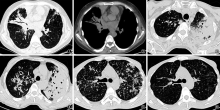
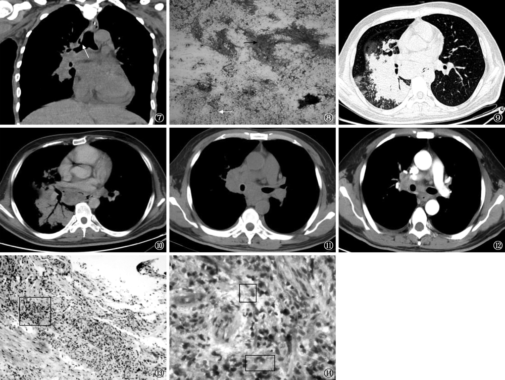
目的: 分析艾滋病(AIDS)合并肺结核患者的胸部CT不典型影像表现,以提高其影像诊断价值。方法: 采用回顾性研究方法,收集2017年1月至2020年1月就诊于首都医科大学附属北京地坛医院经病理、实验室检查证实或临床确诊为AIDS合并肺结核且无其他合并感染的171例患者胸部CT影像资料,分析其病变类型、分布、形态及变化情况。结果: 171例患者中,多种肺结核类型共存117例(68.4%),以继发性肺结核合并胸内淋巴结结核发生率最高[74例(43.3%)]。117例继发性肺结核患者中,37例(31.6%)病灶只分布于好发部位,25例(21.4%)病灶只分布于非好发不典型部位,55例(47.0%)病灶好发部位与非好发部位均有分布;影像征象以支气管播散[85例(72.6%)]、实变[79例(67.5%)]及多发结节[78例(66.7%)]病变为主,其中31例(39.2%)实变病灶表现为类似肺炎的渗出性实变,周围不伴卫星病灶。53例血行播散性肺结核患者中,45例(84.9%)表现为非“三均匀”粟粒结节。107例胸内淋巴结结核患者中,31例(29.0%)病灶破溃侵及临近肺实质、23例(21.5%)病灶融合、5例(4.7%)病变淋巴结有含气征象。结核性胸膜炎和伴发心包积液的发生率均较高[均为36.3%(62/171)]。139例抗结核药物治疗后复查CT平均时间为17d,肺结核病变吸收患者为52例(37.4%),进展患者为58例(41.7%)。结论: AIDS合并肺结核常表现为多种肺结核类型并存;病变广泛,无优势分布;类似肺炎型实变病变比例高;胸内淋巴结结核可有少见含气征象;非“三均匀”的血行播散性肺结核多见;抗结核治疗后病灶短期内变化快。

关键词: 结核,肺, 获得性免疫缺陷综合征, 重叠感染, 放射影像解释,计算机辅助, 诊断显像

Abstract:

Objective: To analyze the chest CT features of AIDS patients complicated with pulmonary tuberculosis (PTB), so as to improve the diagnostic value of imaging. Methods: One hundred and seventy-one AIDS patients only complicated with PTB from Beijing Ditan Hospital, Capital Medical University were retrospectively collected. The diagnosis was confirmed by pathology, laboratory examination or clinical diagnosis from January 2017 to January 2020. The type, distribution, morphology and changes of chest CT were analyzed. Results: Among the 171 patients, 117 (68.4%) had multiple types of PTB, and the incidence of secondary PTB combined with intrathoracic lymph node PTB was the highest (n=74, 43.3%). Of the 117 secondary PTB patients, 37 cases (31.6%) were located at the predilection site, 25 cases (21.4%) were located at the non predilection and atypical sites, and 55 cases (47.0%) were located at both the predilection and non predilection sites. The imaging signs were mainly bronchial dissemination (n=85, 72.6%), consolidation (n=79, 67.5%) and multiple nodules (n=78, 66.7%); 31 cases (39.2%) showed exudative consolidation similar to pneumonia without satellite lesions around. Among 53 cases of hematogenous disseminated PTB, 45 cases (84.9%) showed “three homogeneous” miliary nodules. Among 107 cases of intrathoracic lymph node TB, 31 cases (29.0%) had ulceration and invasion of adjacent pulmonary parenchyma, 23 cases (21.5%) had focal fusion, and 5 cases (4.7%) had gas signs in the lymph nodes. The incidence of TB pleurisy and associated pericardial effusion was high (36.3%, 62/171). The average time of CT re-examination in 139 cases after anti-tuberculosis drug treatment was 17 days, PTB lesions were absorbed in 52 cases (37.4%), while 58 cases (41.7%) had progressed. Conclusion: AIDS complicated with PTB often showed multiple types of PTB. The lesions were extensive and had no dominant distribution; the proportion of consolidation lesions similar to pneumonia was high; intrathoracic lymph nodes TB might have rare signs of gas; non-“three homogeneous” hematogenous disseminated PTB was common; the lesions changed rapidly in a short time after anti-TB treatment.

Key words: Tuberculosis, pulmonary, Acquired immunodeficiency syndrome, Superinfection, Radiographic image interpretation, computer-assisted, Diagnostic imaging

中图分类号: