Email Alert | RSS    帮助

中国防痨杂志 ›› 2022, Vol. 44 ›› Issue (4): 329-335.doi: 10.19982/j.issn.1000-6621.20210713

• 论著 • 上一篇    下一篇

非活动性与活动性肺结核的CT表现分析

李春华1,2, 刘雪艳1, 唐光孝1, 舒伟强1, 王媱1, 王佳男1, 郑娇凤1, 李咏梅2, 吕圣秀1()   

  1. 1重庆市公共卫生医疗救治中心医学影像科,重庆 400036
    2重庆医科大学附属第一医院放射科,重庆 400016
  • 收稿日期:2021-12-21 出版日期:2022-04-10 发布日期:2022-04-06
  • 通信作者: 吕圣秀 E-mail:598341390@qq.com
  • 基金资助:
    重庆市科卫联合医学科研项目(2022MSXM142)

CT features in patients with inactive and active pulmonary tuberculosis

LI Chun-hua1,2, LIU Xue-yan1, TANG Guang-xiao1, SHU Wei-qiang1, WANG Yao1, WANG Jia-nan1, ZHENG Jiao-feng1, LI Yong-mei2, LYU Sheng-xiu1()   

  1. 1Department of Radiology, Chongqing Public Health Medical Center, Chongqing 400036, China
    2Department of Radiology, the First Affiliated Hospital of Chongqing Medical University,Chongqing 400016, China
  • Received:2021-12-21 Online:2022-04-10 Published:2022-04-06
  • Contact: LYU Sheng-xiu E-mail:598341390@qq.com
  • Supported by:
    Chongqing Medical Scientific Research Project (Joint Project of Chongqing Health Commission and Science and Technology Bureau)(2022MSXM142)

摘要:

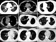
目的: 对比分析非活动性肺结核与活动性肺结核CT表现。方法: 回顾性搜集2020年8月至2021年7月重庆市公共卫生医疗救治中心诊治的181例非活动性肺结核患者(非活动组)和 166例活动性肺结核患者(活动组),分析两组患者的胸部CT表现。结果: 非活动组病变累及1叶者[19.9%(36/181)]和2叶者[25.4%(46/181)]均明显多于活动组[分别为10.8%(18/166)和13.3%(22/166)](χ2值分别为5.392、8.128,P值分别为0.020、0.004),而5叶均受累者[21.5%(39/181)]明显低于活动组[48.2%(80/166)](χ2=27.283,P=0.000);病变在右肺中叶、下叶及左肺下叶者[分别为38.7%(70/181)、45.3%(82/181)、46.4%(84/181)]均明显少于活动组[69.9%(116/166)、77.1%(128/166)、68.7%(114/166)](χ2值分别为33.903、36.657、17.520,P值均为0.000)。非活动组中小片状实变影、干酪性病变、空洞、胸腔积液、纵隔淋巴结肿大和树芽征等CT表现的发生率[分别为22.1%(40/181)、0.6%(1/181)、16.6%(30/181)、0.6%(1/181)、18.8%(34/181)、18.2%(33/181)]均明显低于活动组[分别为80.1%(133/166)、7.2%(12/166)、27.1%(45/166)、31.9%(53/166)、53.6%(89/166)、66.9%(111/166)](χ2值分别为116.598、10.703、5.671、64.868、45.906、84.365,P值分别为0.000、0.001、0.017、0.000、0.000、0.000),但边缘清楚的支气管扩张、薄壁空洞、胸膜钙化、钙化结节、硬结性病变、纤维条索影的发生率[分别为61.3%(111/181)、12.2%(22/181)、10.5%(19/181)、34.8%(63/181)、37.0%(67/181)、91.7%(166/181)]均明显高于活动组[分别为44.0%(73/166)、4.2%(7/166)、3.6%(6/166)、16.9%(28/166)、0.6%(1/166)、27.7%(46/166)](χ2值分别为10.464、7.124、6.135、14.403、70.576、149.222,P值分别为0.001、0.008、0.013、0.000、0.000、0.000)。结论: 非活动性肺结核病变分布较少累及下叶,CT表现以纤维条索影、边缘清楚的支气管扩张、硬结性病变、结节和胸膜钙化更常见。CT检查对肺结核活动性的判断具有重要辅助诊断作用。

关键词: 结核,肺, 诊断, 体层摄影术,X线计算机

Abstract:

Objective: To investigate the CT features in patients with inactive pulmonary tuberculosis and active pulmonary tuberculosis. Methods: All of 181 patients with inactive pulmonary tuberculosis (inactive group) and 166 patients with active pulmonary tuberculosis (active group) diagnosed and treated in Chongqing Public Health Medical Center from August 2020 to July 2021 were retrospectively collected, and CT manifestations in the two groups were analyzed. Results: The percentages of lesions involving one lobe (19.9% (36/181)) and two lobes (25.4% (46/181)) in the inactive group were significantly higher than that in the active group (10.8% (18/166) and 13.3% (22/166), respectively)(χ2=5.392 and 8.128 respectively, and P=0.020 and 0.004, respectively). The percentage of lesions involving 5 lobes (21.5% (39/181)) was significantly lower than that in the active group (48.2% (80/166))(χ 2=27.283, P=0.000). The percentages of lesions in the right middle lobe and lower lobe as well as the left lower lobe (38.7% (70/181), 45.3% (82/181) and 46.4% (84/181), respectively) were also significantly lower than those in the active group (69.9% (116/166), 77.1% (128/166) and 68.7% (114/166))(χ 2=33.903, 36.657, 17.520; Ps=0.000). CT findings: the incidences of patchy consolidation (22.1%, 40/181), caseous lesions (0.6%, 1/181), cavity (16.6%, 30/181), pleural effusion (0.6%, 1/181), lymphadenopathy (18.8%, 34/181) and tree-in-bud signs (18.2%, 33/181) in the inactive group were significantly lower than those in the active group (80.1% (133/166), 7.2% (12/166), 27.1% (45/166), 31.9% (53/166), 53.6% (89/166), 66.9% (111/166))(χ 2=116.598, 10.703, 5.671, 64.868, 45.906 and 84.365, respectively, and P=0.000, 0.001, 0.017, 0.000, 0.000, 0.000, respectively). The incidences of bronchiectasis (61.3%, 111/181), parenchyma cavity (12.2%, 22/181), pleural calcification (10.5%, 19/181), calcified nodules (34.8%, 63/181), sclerosis (37.0%, 67/181) and fibrosclerosis (91.7%, 166/181) in the inactive group were significantly higher than those in the active group (44.0% (73/166), 4.2% (7/166), 3.6% (6/166), 16.9% (28/166), 0.6% (1/166), 27.7% (46/166))(χ 2=10.464, 7.124, 6.135, 14.403, 70.576 and 149.222 respectively, and P=0.001, 0.008, 0.013, 0.000, 0.000 and 0.000, respectively). Conclusion: The lesions of inactive pulmonary tuberculosis in the lower lobe are less distributed, and fibrosclerosis, bronchiectasis, sclerosis, calcified nodules and pleural calcification are more common signs on CT. CT scanning plays an important role in assessing the activity of pulmonary tuberculosis.

Key words: Tuberculosis,pulmonary, Diagnosis, Tomography,X-ray computed

中图分类号: